XenMD – Xenopus Modelling Disease XenMD for Patients and Families About Us
XenMD Overview
XenMD provides a diagnostic support service to clinicians with patients presenting in England with a rare genetic disease and complex symptoms. All patients are referred by clinicians (we do not accept private referrals) following sequencing analysis, when a diagnosis cannot be found based on computer or cell-based research. Here a frog model, Xenopus tropicalis, is used to better understand changes that have occurred in the genome of each of these patients by modelling the exact changes in the frog. This information is then fed back to the clinical team caring for each patient.
Genetics
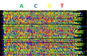
 The make up for our bodies is largely dictated by a 1.8 m long string of four chemical letters (A,T,G and C), this is our genome and together the chemical letters strung together are called DNA. The DNA is effectively a plan for making proteins, which are the “doing molecules” in our bodies; the globin proteins are “encoded” by the globin genes for example, they make up haemoglobin which carries oxygen around our body in red blood cells. If a chemical letter is very unusual (a rare variant) in someone’s globin gene then it may make no difference to how well their globin protein works at all (a benign variant) or it might cause a rare genetic disease like thalassaemia or sickle cell (a pathogenic variant). XenMD is about gene variants that are previously unknown but that DNA sequencing of a patient and their parents, followed by computational analysis, suggests but does not show is responsible for a disease; these are variants of uncertain significance.
How well do you know a Xenopus frog?
 The frogs we use were native to sub-Saharan Africa, where they would typically be found in a pond with slow moving water. Xenopus frogs are unusual looking. They have flattened bodies with small heads, no eyelids, muscular hind limbs, fully webbed toes with claws to rip apart food and help shed their skin. Xenopus have a mottled skin normally greenish, grey in colour on their back with a lighter coloured underside. The frogs’ skin gives them camouflage from predators and it can be used in captivity to identify individual animals. Female Xenopus frogs are about 33% bigger than males. Sexually mature males develop ‘black gloves’ on their front legs. These gloves are like velcro and they are used to hold onto the females abdomen during mating, in a position called amplexus. Their gloves are shed shortly after mating. Xenopus are very relaxed and calm most of the time, they like to hide in tunnels or chill out on top of lily pads and they communicate using sounds. Frogs feed using their front limbs to shovel food in, in what is often described as a feeding frenzy. These frogs in the wild would be nocturnal hunters and they eat all types of animals (including each other, so we keep them in groups where frogs are the same size!).
How similar are frogs and humans?
 Frogs were first used in the clinic in the 1940s to 1970s when they were used as a pregnancy test. The urine of pregnant women was injected into the frog and if the frog subsequently laid eggs, the women was pregnant. This is a technique still used in research today; egg laying is induced in animals in captivity all year round using hormone injections. Using frogs for diagnosis is now coming back into medicine because we can alter frogs’ genomes to test whether a variant in a patient’s genome causes their disease; if the gene variant is causative the genome change in the tadpole will give it some of the patient’s symptoms (called a phenotype). This process is shown in the flow diagram below. To do this it is of course important that the frog is similar to humans both as an animal and genetically. Frogs are tetrapods (four legged) like humans and their general body plan is like ours as adults, but we do many of the phenotype tests as tadpoles when they lack lungs and legs. We know that many of the basic biological processes in the frog are the same as in humans from more than 70 years of work from around the world in which many of the developmental, cellular and molecular processes that happen in our bodies were first discovered using frogs. This, and many successful experiments already completed, make us confident that frogs are good for modelling human diseases.

In relation to genetics, 90% of known human disease-causing genes are found in the frog species we use. Like the human’s the frog genome is very accurately sequenced and we know how it relates to ours. There are three really important genetic things to understand about why we use frogs: first, it is rare for us to find a disease gene that is too dissimilar between frogs and humans to model the gene variant; second these frogs are properly diploid, they have 2 copies of each gene like us, which makes it much more straightforward to mimic a gene change; finally the genome is largely structured the same in frogs and humans, having a similar gene order makes it easier to work out exactly how to mimic the gene variants.
These similarities allow us to use frogs for experiments that would be impossible, unethical or cost-prohibitive in people or mammals. This does not mean that occasionally frogs are not a good model, and in that case we have a mechanism in place to refer these diseases to a mouse group specialising in similar studies. Finallywe do this work to the very highest ethical and welfare standards that we can, often beyond those set in law. If there are alternatives such as cultured cells we will not use animals, and we only make the disease models if clinicians are sure it is necessary to test the gene variant-disease link and thus important for patients and their families.
Additional Resources


