Line name: Xla.Tg(mlc3:eGFP)Mohun
Synonyms: MLC1v:EGFP
RRID: EXRC_21
EXRC line: #21
Non-standard MTA required? No
Transgene description: 8Kb of X. laevis mlc3 ( synonym = MLC1v ) gene driving expression of enhanced GFP (eGFP). Promoter sequence is NCBI AY289208. Plasmid linearised with PmeI was inserted. Select best expressing animals at NF stage 37, monitor to NF stage 45.
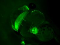
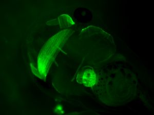
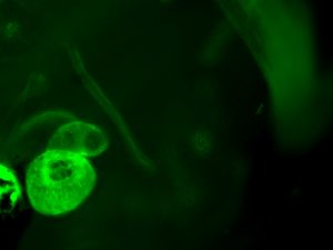
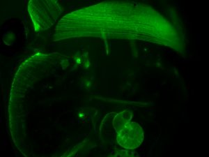
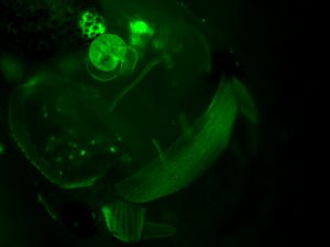
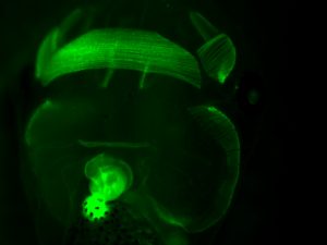
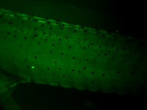
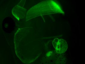
Phenotype: GFP expression in heart myocardium.
Source lab: Mohun lab
Publication: Smith, S. et al (2005) “ The MLC1v Gene Provides a Transgenic Marker of Myocardium Formation Within Developing Chambers of the Xenopus Heart. Dev. Dyn. 232:1003–1012
Species: laevis
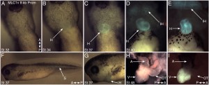
Fig 1, The onset of ventricular cardiac expression for the MLC1v promoter detected in MLC1v::EGFP transgenic embryos.
A-E: Ventral views of the heart-forming region of a single MLC1v::EGFP animal at stage 32 (A), stage 34 (B), stage 37 (C), stage 40 (D), and stage 43 (E). F, G: Left lateral views of the same tadpole at stage 37, depicting the whole animal (F) and detail of the head (G). H, I: The heart, dissected from a stage 48 transgenic tadpole (H, bright field image) reveals its chamber myocardium-restricted EGFP fluorescence (I, dark field image). H, heart; IH, interhyoid facial muscle; S, somites; A, atria; V, ventricle; OT, outflow tract.
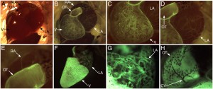
Fig 2, MLC1v promoter activity observed in MLC1v::EGFP transgenic frog hearts.
A-E: The heart of an 80 day old, recently-metamorphosed froglet. Terminal anaesthesia was used to stop the animal’s heart beat and its pericardium removed by dissection to reveal the cardiac chambers. The atria are filled with blood, ventricle part-filled. A, B: Bright-, and dark field views of the entire heart. C-E: High magnification views of the single ventricle (C), left atrium (D), and right atrium and proximal outflow tract (E). F-H: The heart, dissected from a 39 week old juvenile frog, showing bright EGFP fluorescence in the ventricle and some myocardial fibres of the atrial chambers (F). High magnification views of the left atrium (G) and proximal outflow tract (H). A coronary vessel in the outflow tract can be seen as a dark silhouette in (H). Distinct MLC1v-expressing myocardial fibre morphologies are observed in the different compartments of the heart. V, ventricle; LA, left atrium; RA, right atrium; OT, outflow tract; CV, coronary vessel.
Copyright Feb 19th 2008
Stuart Smith asserts his rights as the owner of each of the images and movies described below.
The MLC1v::EGFP images have been published previously so Developmental Dynamics holds the copyright.
The dark field images that portray the fluorescence signal from GFP have had their input/output levels for the red, green and blue colour channels adjusted individually in Adobe Photoshop. This approach enables the silhouette of the animal to be observed more clearly, while giving the EGFP fluorescence a false, blue-green colour.
MLC1v Promoter +Flanking Sites Sequence
MLC1v Promoter-AY289208 Sequence
Line information can also be found in Xenbase.
