Line name: Xla.Tg(nkx2-5b:GFP)Mohun
Synonyms: Nkx2-5:GFP
RRID: EXRC_0019
EXRC line: #19
Non-standard MTA required: No
Transgene description:8 kb of promoter of the X. laevis nkx2-5 gene driving expression of GFP (variant mt5). Mohun Lab Plasmid #2147. Contact Tim Mohun for the sequence as it differs from the published NCBI AF283102. The promoter of the second laevis Nkx2-5 allele was used here. It proved stronger than the original published one (Sparrow et al 2000 Dev. Biol. 227, p65 to p79.)
Source lab: Mohun lab
Phenotype:GFP expression in heart precursors
Publication:
Sparrow, D. et al (2000) “Regulation of the tinman Homologues in Xenopus Embryos”, Dev. Biol. 227, 65–79.
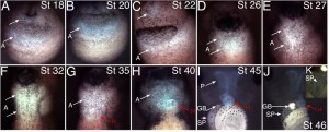
Fig. 1. Heart field expression of the Nkx2-5::GFP transgenic line in Xenopus laevis.
A-C: Fluorescence from the Nkx2-5::GFP transgene is first observed during late neurula and early tailbud stages of development. A: Anterior ventral view of a stage 18 embryo. B: Stage 20 embryo. C: Ventral view of the anterior portion of a stage 22 embryo. The GFP fluorescence that is viewed on both anterior and posterior sides of the cement gland (A, C-arrows) actually forms a contiguous domain of pharyngeal GFP expression. D, E: The shape of the GFP domain changes as the tailbud stage embryo elongates along the anterior-posterior axis. D: Ventral view of the heart-forming region at stage 26. E: Stage 27. When viewed from the ventral side, the fluorescence appears brightest in a posterior part of the domain where the cardiac mesoderm and underlying pharyngeal endoderm both express GFP. F-K: During tadpole development, it becomes easier to discriminate between the cardiac mesoderm and pharyngeal endoderm components of the transgene activity and the heart commences beating. F: Stage 32. G: Stage 35. H: Stage 40. I: Stage 45. J: Stage 46. K: Detail view of the forming spleen in a stage 46 animal, an organ that expresses Nkx2-5 mRNA and also shows transgene activity. Melanocyte pigment formation was inhibited in this tadpole to give clearer photography. White arrows indicate the extent of the pharyngeal expression domain while red arrows point to the heart. A, anterior domain of GFP; H, heart; P, pharynx; GB, gall bladder; SP, spleen.
Copyright Feb 19th 2008
Stuart Smith asserts his rights as the owner of each of the images and movies described below (simply because I haven’t published any of them elsewhere).
The dark field images that portray the fluorescence signal from GFP have had their input/output levels for the red, green and blue colour channels adjusted individually in Adobe Photoshop. This approach enables the silhouette of the animal to be observed more clearly, while giving the EGFP fluorescence a false, blue-green colour.
Line information can also be found in Xenbase.


